Visualizing CNS Infections: A Recent Collaboration
Bringing Clarity to CNS Infections Through Illustration
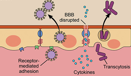
I recently had the opportunity to illustrate seven full-color neuroanatomical and vascular images for the chapter “Anatomical Organization of the Central Nervous System (CNS)” in Elsevier’s new 2025 volume, Neurobiology of Infectious Diseases. Authored by neurologists Guadalupe Ortiz, Carlos Martinez-Menendez, Kristofer Harris, Miriam Hinojosa, and Paul Schulz, the chapter provides an in-depth look at the CNS in the context of mechanisms and pathways to neurological infections.
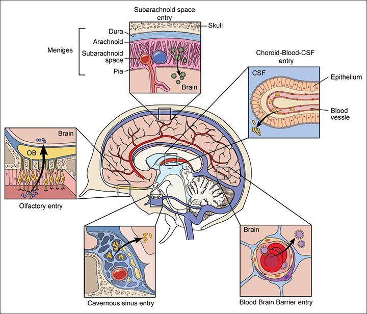
My illustrations were designed to clarify key structures involved in brain and spinal cord anatomy, with a focus on the pathways and regions most affected by infectious disease. These visuals support both teaching and clinical understanding, helping to bridge the gap between dense neurological content and accessible medical communication. The illustrations were created in Adobe Illustrator in a simple but detailed line and color style.
Collaborating with this expert team was both challenging and rewarding, especially in a field where precise visuals can enhance both comprehension and impact. It’s always fulfilling to contribute to medical education through meaningful, evidence-based illustration.
You can explore more of this kind of work right here in my portfolio—or check out the book if you’re diving deep into the world of neuroinfectious disease.